Tibia and tibia diagram. The skeleton of the free part of the lower limb. Fracture consequences and prevention
The shin bones are part of the peripheral skeleton, which connects the bones of the chest and pelvic limbs. The tibia and fibula form the tibia. Injuries to these parts of the skeleton immobilize a person for a long time and pose a threat to his health.
The structure of the tibia
As we have already found out, the tibia and fibula form the tibia, and are located in its inner part. If we put our hand on the front of the leg (below the knee), then we will immediately rest on the tibia. And on the outside of the lower leg is the fibula, which cannot be touched, since it is located in the thickness of the muscles. Therefore, these two bones are connected to each other and form an ankle joint on one side and a knee joint on the other. Thus, their structure determines the mobility and functionality of the lower limbs.
Tibia
The tibia is located closer to the center in relation to the lesser bone. It is a tubular long bone, which is equipped with two epiphyses and a body. Its body consists of three edges, which are triangular in shape:
- front;
- interosseous;
- medial.
These edges have three surfaces:
- back;
- medial;
- lateral.
The upper epiphysis, together with the patella, forms the knee joint. The lower part articulates with the talus and forms the ankle. The tibia is the most massive and stable bone in the human skeleton. She experiences the greatest stress when a person is standing, running or walking fast. In addition, this bone is very light because it has a microscopic structure, it is penetrated by multiple vessels and nerve endings.
Tibia
Located on the outer (lateral) side of the lower leg. It is also a long tubular bone, but much smaller in shape and thickness. Consists of two epiphyses: upper and lower. The upper one fits into the knee joint, and the lower one into the ankle. As part of the ankle joint, it is called lateral (external ankle). Its main function is to stabilize the ankle joint. However, it practically does not bear any load, but is the place of muscle attachment.

Has three surfaces:
- back;
- medial;
- lateral.
These surfaces are separated by three ridges.
Trauma
Injury to the lower extremities occurs due to the heavy load on the joints that they experience every day when walking and moving. Injuries to the lower leg usually damage both bones.
In addition, in some cases, this load increases:
- if you are overweight or obese;
- congenital anomalies of the skeletal system (in this case, the lower extremities);
- with a weak muscular apparatus;
- in violation of coordination of movements.

In these cases, the bones cannot cope with the load that is placed on them, which leads to injury. Such damage occurs for different reasons and, depending on this, differ in nature and severity. For example, with direct damage to the bone, fragments of one type are observed, and with indirect injury, of another type.
Causes of damage to the shin bones:
- swipe;
- car crashes;
- falling from a height;
- work injuries;
- excessive physical activity (for example, during professional sports).
Fracture classification
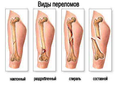
Injuries to the lower leg usually damage both bones. Fractures of the body of the tibia bones are almost always accompanied by the displacement of bone fragments. They are of the following types:
- Transverse. If only the tibia is fractured, then there is a stable damage to the bones without displacement of the fragments. If a small bone is damaged, then the instability of the fragments is noted.
- Helical. Observed under the influence of a twisting force, the damage is unstable and has a spiral shape.
- Oblique. Typically occur at an angle. The damage is unstable, with a tendency to increase displacement.
- Comminuted. They are characterized by severe instability and the formation of more than three bone fragments.
In addition to this classification, there are open and closed fractures. With closed fractures, there is no violation of the integrity of the skin.
When open, the skin is damaged, and the broken fragments communicate with the external environment. This type of damage is also dangerous because the resulting wound can become infected.
Such injuries are far from uncommon; they occur in both adults and children. You do not need to have special medical knowledge to understand that trauma to this anatomical segment is very dangerous and can lead to serious consequences.
Injuries involving damage to both bones are especially dangerous. Indeed, in this case, a person will be completely immobilized and a long rehabilitation. Displaced fractures are possible, which also require a long recovery period.
Fracture symptoms
Symptoms are characterized by severe, sharp pain, rapidly increasing swelling, bruising and bruising, and obvious shortening of the injured limb. The victim cannot not only walk, it is impossible to lean on and simply stand on the injured limb. As a rule, such fractures always occur with displacement of fragments. The leg may take the wrong position and be turned in a certain direction: inward or outward (in relation to the knee). With an open fracture, skin damage is observed, through which bone fragments are visible.
The diagnosis is made by X-ray examination, since the clinical picture alone is not enough. The study of the radiograph allows you to determine the number of fragments and the degree of their displacement, the presence of a fracture of both bones or only one of them, as well as the integrity of the knee and ankle joints. They also determine the integrity of blood vessels and nerves. For this, the victim is sent for consultation to narrow specialists.
Treatment and first aid
The provision of first aid can affect the further treatment and rehabilitation of the victim. First of all, he is given analgesics and anti-shock therapy (in the presence of multiple injuries). The shin is immobilized using a splint. Any object that is at hand (plywood, skis, boards) can act as a tire. When applying a splint, it is very important that the lower part of the splint covers the ankle joint, and the upper part ends at the upper part of the thigh.

For an open fracture, a tourniquet should be placed just above the wound to stop the bleeding. It is imperative to treat an open wound with iodine, alcohol, brilliant green, or just rinse with water if disinfectants are not at hand. All of these steps are necessary to minimize wound infection.
Conservative treatment
Treatment in a medical facility can be both conservative and surgical. Treatment tactics depend on the degree and level of damage. In case of damage that is stable and without displacement (which happens extremely rarely), a plaster cast is applied. For other types of damage, skeletal traction is used. The essence of this treatment is that a metal needle is passed through the heel bone, and a splint is applied to the leg.
This treatment involves two scenarios. First, conservative treatment involves traction for 4 weeks, during which the bone fragments are fixed in the correct position. When a callus appears, the skeletal traction is removed and a plaster cast is applied for another two months. Secondly, after removing the bandage, the patient is prescribed rehabilitation: physiotherapeutic procedures, massage and therapeutic exercises.
Operative treatment
Surgical treatment is indicated for multiple fractures that are difficult to restore to the correct anatomical position with conventional conservative treatment. Surgical treatment involves the use of a variety of metal structures - plates, pins, rods. In addition, with such injuries, the use of the Ilizarov apparatus is indicated. The device allows you to restore the natural location of the fragments and their rapid fusion. It is used in the most difficult cases - for fragmented fractures with the formation of a bone defect. The period of bone healing is approximately 4-6 months. The rate of recovery is individual and depends on the degree of damage and the complexity of the injury.
The tibia is part of the lower leg skeleton. Its damage can permanently deprive a person of the ability to move. If the bones do not heal or are not connected correctly, surgery may be required.
Location
The shin is where the shin bone is located. It is in two parts and is located at the bottom of the leg. The tibia (tibia) is located medially. It is long, has a 3-sided body and two pineal glands. The upper end of the tibia is involved in the formation of the knee joint. The tibia bone is the strongest in the human skeleton. The tibia can withstand a maximum load of 1650 kilograms.
The fibula (MBC) is less massive and is located laterally. It is long and tubular, attaches to the large and restricts the ankle. Fractures and injuries of the MBC are rare.
Description of BBK
The largest component of the tibia is called the tibia, its anatomy has one feature. Its second, but separate, half adjoins the LBC. This is the small bone of the tibia. The tibia and fibula are attached to the hip joints and patella. An ankle is formed below and adjoins the talus.
The front edge of the tibia looks like a pointed crest. From above it is bumpy. There is a small connecting cartilage between the tibia. The surface of the tibia is convex and can be felt even through the skin. The lateral part is concave, the posterior part is flat, with a soleus muscle. Below is the feeding hole.
The proximal pineal gland is slightly enlarged. Its sides are called condyles. Outside the lateral is an articular flat surface. At the top of the proximal pineal gland there is a small eminence with two tubercles. The distal pineal gland is quadrangular. The lateral surface has a peroneal notch. Behind the pineal gland is the ankle groove.
LBC fractures
With injuries to the tibia, where it is located, pain appears. This may indicate her fracture. The latter can have several varieties. Fractures of the shin bones are oblique and transverse. They also distinguish between comminuted and fragmentary.
Intra-articular fractures may appear in the condyles or medial malleolus. Most often this occurs due to twisting of the lower leg with a fixed foot. This is manifested in the fact that a person has pain in the tibia. Ankle fracture often occurs after a sharp turn of the foot.
Bone fracture symptoms
Even small cracks in the bones are negative. Fractures feel much more acute. They are detected quickly when the tibia bone hurts when walking - this may indicate a violation of its integrity. Unpleasant sensations arise when feeling the feet. Severe pain is immediately felt at the fracture site.
If the bone fragments turned out to be displaced, then the lower leg is deformed and the axis of the limb changes. Swelling appears on the leg. The limb cannot bear any load. After surgical treatment of the deformed tibia, a person can stand on the sore leg the next day after the operation.
 When the proximal part is injured, acute pain occurs, which intensifies when the limb is felt. The leg becomes shorter, it is impossible to step on it, it does not bend at the knee. I can't even move the affected limb.
The first sign of diaphyseal fractures is the appearance of extensive hematomas. They are formed due to subcutaneous hemorrhage in soft tissues. Sometimes a state of shock appears. With such a fracture, a person cannot move, he is tormented by severe pain. Fragment fractures are very rare, but still occur. In this case, swelling and pain immediately appear.
Why does the large tibia bone hurt? This can be with a simultaneous fracture and MBC. As a result of trauma to both shin bones, treatment is greatly complicated. With such a fracture, if displacement is observed, it is impossible to carry out the usual reduction.
Cyst
When the shin bone hurts, it may mean the appearance of a cyst. This is an ailment when a thickening appears in half of the tissue. Cysts are a manifestation of a dystrophic process.
At the heart of the thickening is impaired blood circulation and the active activity of lysosomal enzymes, which lead to a decrease in collagen and other useful substances and proteins. A cyst refers to a neoplasm that can be both benign and malignant.
They are found when the shin bone on the leg begins to hurt. The cyst is aneurysmal or solitary. It develops over a long period of time. A solitary cyst is most often found in adolescents. Aneurysmal neoplasm appears suddenly. Basically, such a cyst appears after an injury or bone fracture.
Pain in the lower leg and her bones
Calf pain can have various causes. For example, from excessive training, when the shin bone begins to hurt after running. It can become more fragile if there is a lack of calcium, magnesium and other essential elements in the body. They are often washed out when a person uses diuretics.
When the shin bone in front hurts, it may be due to joint disease or excessive stress that the legs suddenly felt after a long stagnant period. The causes of negative sensations can be inflammation or an infection that affects the bone tissue. Very rarely, a malignant tumor can appear on the bone.
MBC fracture
 Trauma or fracture of the MBC may result from damage to the head or neck. This rarely happens. Most often, such a fracture is combined with other injuries of the lower leg. A person immediately feels severe pain in the knee. Nevertheless, the leg is able to bend and unbend.
The bad news is that in the MBC, the upper section can cause very serious complications. They are due to nerve damage and dysfunction. This provokes additional complications, up to the complete immobilization of the limbs. For MBC fractures, conservative treatment is performed. But if complications arise, a surgical operation is performed.
Complications after fractures
Complications after fractures can occur most often due to an untimely visit to the surgeon or after improper treatment. But often it is not doctors who are to blame for the complications, but the individual characteristics of the organism (intolerance to certain drugs, low content of calcium in the tissues, etc.).
Complications can manifest in different ways. Incorrect fusion of the tibia where the fracture was. Fat embolism occurs, blood supply to internal organs is disrupted. After the fusion of the bones, complete immobilization of the leg or knee occurs. Deforming osteoarthritis may begin in them. During healing due to a bone defect, a false joint is observed. Deformation of the leg occurs.
A tibia fracture is the most common complication. Often they begin due to the forced long immobilization of the leg. But thanks to modern means and technology, most of the negative consequences have become possible to avoid.
Fracture treatment
 Fractures are most often treated on an outpatient basis. A plaster cast is applied to the limb. In addition, the limb can be additionally secured with special devices. In order to calculate in time how much the tibia bone grows together, you need to start from the moment the leg is fixed.
After applying the plaster, a ten-day bed rest is prescribed. Then the person is allowed to walk a little and lightly step on the foot. Most often, the bones are completely healed within five weeks. A complex fracture of the tibia may require inpatient treatment. In this case, fusion occurs within two months.
If it is revealed that the tibia bone (its photo is in this article) is broken with displacement and the presence of fragments, then the fragments are repositioned first. The operation takes place under local anesthesia. After that, the cast is applied to the entire leg. Treatment of condylar injuries and fractures is carried out using osteosynthesis and traction. In this case, the healing of the leg takes two to four months. The main thing is not to delay a visit to a specialist and start treatment on time.
Cure arthrosis without medication? It's possible!
Get a free book "17 recipes for delicious and inexpensive dishes for the health of the spine and joints" and begin to recover without effort!
Get the book
Ankle joint and its diseases
The ankle is one of the most vulnerable joints in the human body. Its damage often leads to complete immobilization of a person. It connects the foot to the lower leg. For normal walking, it is necessary that he be healthy and fully perform his functions.
The ankle joint allows any movement of the foot. The anatomy of the ankle is quite complex. It consists of several bones that are connected by cartilage formations and muscle ligaments.
Anatomical features
The distribution of the pressure of a person's body weight over the surface of the foot is provided by a nominal ankle, which accounts for the load of the entire person's weight. The upper anatomical border of the ankle runs along a conditional line 7-8 cm above the medial ankle (visible projection from the inside). The border between the foot and the joint is the line between the lateral and medial ankles. The lateral ankle is on the back of the medial.
The joint is divided into inner, outer, anterior and posterior sections. The back of the foot is the forefoot. In the area of \u200b\u200bthe Achilles tendon is the posterior section. In the area of \u200b\u200bthe medial and lateral malleolus - the inner and outer sections, respectively.
Joint bones
The ankle joint connects the fibula and tibia with the supracal bone - the talus or foot bone. The process of the foot bone enters the socket between the lower ends of the fibula and tibia. The ankle joint is formed around this joint. Several elements are distinguished in this basis:
- the inner ankle is the lower (distal) edge of the tibia;
- outer ankle - the edge of the fibula;
- the distal surface of the tibia.
The external ankle at the back has a depression in which the tendons are fixed, suitable for the muscles of the peroneal muscles - long and short. The fascia (connective tissue sheaths), together with the lateral articular ligaments, are attached to the outside of the outer ankle. Fasciae are formed from protective sheaths that cover tendons, blood vessels, and nerve fibers.
The ankle joint has a so-called gap, which is formed on its inner surface by the upper side of the talus and hyaline cartilage.
Ankle appearance
The structure of the ankle joint is not difficult to imagine. The surface of the lower edge of the tibia looks like an arc. The inner side of this arch has a process. Below on the tibia there are processes in front and behind. They are called the front and back ankles. The fibular notch on the tibial is on the outside. There are bumps on the sides of this notch. The outer malleolus is partially located in the peroneal notch. She and the peroneal notch together create tibiofibular syndesmosis. For the full functioning of the joint, its healthy condition is very important.
The front is smaller than the back. The joint surface is divided into inner and outer by a bony ridge.
The anterior and posterior tubercles of the articular surface form the inner ankle. They are separated from each other by a fossa. The anterior tubercle is larger than the posterior one. The deltoid ligament and fascia are attached to the ankle from the inside without articular surfaces. The opposite surface (from the outside) is covered with cartilage.
The calcaneus and shin bones are connected by the talus, which consists of the head, neck, block and body. The talus block provides connection to the lower leg. Between the distal parts of the fibula and tibia, a “fork” is formed, in which the talus block is located. The block is convex on the upper side, along it there is a depression, into which the crest of the distal epiphysis of the tibia enters.
The block is slightly wider at the front. This part goes into the neck and head. On the back there is a small tubercle with a groove along which the flexor of the thumb passes.
Joint muscles
Behind and outside of the ankle, there are muscles that provide flexion of the foot. These include:
- long flexors of the toes;
- posterior tibial;
- plantar;
- triceps muscle of the leg.
In the anterior part of the ankle, there are muscles that provide extension:
- anterior tibial;
- extensors of the toes.
The short long bone and the third fibula are the muscles that move the ankle outward (pronators). Inward movement is provided by the instep supports - the long extensor of the thumb and the anterior tibial muscle.
Ankle ligaments
Normal function and movement in the joint is provided by ligaments, which also hold the bony elements of the joint in place. The most powerful ankle ligament is the deltoid. It connects the talus, calcaneus, and navicular bones (foot) to the inner ankle.
A powerful formation is the ligamentous apparatus of the tibiofibular syndesmosis. The shin bones are held together by the interosseous ligament, which is an extension of the interosseous membrane. The interosseous ligament passes into the posterior lower ligament, which keeps the joint from turning in too much. The anterior inferior tibiofibular ligament restrains from too strong an outward rotation. It is located between the peroneal notch, which is located on the surface of the tibia and the outer malleolus. In addition, the transverse ligament located under the tibiofibular ligament prevents excessive rotation of the foot outward.
Blood vessels
Tissue nutrition is provided by the peroneal, anterior and posterior tibial arteries. In the region of the joint capsule, ankles and ligaments, the vasculature diverges from these arteries, as the arteries branch out.
The outflow of venous blood occurs along the external and internal networks, which converge into the anterior and posterior tibial veins, the small and large saphenous veins. The venous vessels are connected into a single network by anastomoses.
Ankle functions
The ankle can perform movements around its axis and along an axis passing through a point in front of the outer ankle. Its own axis passes through the center of the inner one. Movement along these axes is possible with an amplitude of 60-90 degrees.
How does ankle pain manifest?
When ankle pain occurs, it is usually difficult for a person to walk. The ankles are swollen, and the skin may turn blue in the affected area. It becomes almost impossible to step on the foot due to a significant increase in pain in the ankle, which loses its ability to support a person's weight.
If the ankle is affected, pain can radiate to the knee or lower leg. Most athletes are at risk for pain in the ankle joint, since when playing football, tennis, volleyball, hockey and other mobile sports, the joints of the legs have a significant load.
There are several of the most common injuries that cause pain in the ankle area. These include injuries - dislocations, subluxations, fractures, etc. The ankle is one of the most susceptible to joint injuries. Every person knows the unpleasant feeling that arises if you twist your leg.
Ankle fracture
The ankle is the area that fractures more often than most bones in the human body. The fracture is usually caused by a sharp and too fast movement of the ankle inward or outward. Often an ankle fracture is accompanied by a sprain of the ankle ligaments. Fractures and other injuries of the ankle are more susceptible to people with weak ligaments. With injuries of the ankle, the joint area swells, severe pain does not allow to stand on the leg.
Tarsal Tunnel Syndrome
This pathology is a neuropathy associated with damage to the posterior tibial nerve. The nerve contracts as if passing through a tunnel. In this case, a person feels a tingling sensation and soreness of the ankle joint. The same sensations can extend to the legs. Feel cold or hot around the ankle and feet.
Tendonitis
With this disease, inflammation of the Achilles tendon occurs. Tendinitis often causes complications such as tendon rupture or arthritis. If pain occurs while running or walking, swelling or pain in the ankle joint, Achilles tendonitis may be suspected. You cannot start his treatment, as this is fraught with frequently recurring injuries, especially for people who often and often walk, run, jump.
Ankle arthritis
The most common ankle disorder is arthritis. Depending on the type of arthritis, the causes may vary, but the most common and common are:
- Infection of the joint with pathogenic bacteria. It can be gonococci, chlamydia, pale spirochetes. In this case, we are talking about a specific form of the disease. The nonspecific form occurs as a secondary disease after influenza or furunculosis.
- Gout. Due to metabolic disorders in the body, the ankle joint can also be affected.
- Immune system disorders. The body can recognize the cells of the articular tissue as foreign and begin to attack them.
- Injury and mechanical damage.
The factors that provoke the development of the disease can be the following:
- wearing uncomfortable shoes;
- flat feet;
- hormonal disorders;
- disruptions in metabolism;
- strong professional loads (mainly for athletes);
- severe hypothermia;
- excess weight;
- hereditary predisposition;
- unhealthy Lifestyle;
- allergies and lowered immunity.
Arthritis is treated conservatively or surgically. With the bacterial form of the disease, antibiotic therapy is required. It is important to follow a special diet to relieve pain and disease. It is necessary to exclude nightshades, canned food and smoked meats from the diet; salt intake must be minimized. To relieve inflammation, NSAIDs (Diclofenac, Voltaren, Aspirin) are prescribed. Painkillers help relieve the patient's condition. It is recommended to take vitamins and dietary supplements to improve metabolism, further relieve inflammation and speedily restore cartilage tissue.
The tibia is a large and long bone of the lower leg. The bone consists of a body and two epiphyses - the lower distal and the upper proximal.
The structure of the tibia
The body of the bone has a triangular shape with three edges - anterior, medial and interosseous, and three surfaces - medial, posterior and lateral.
The anterior edge of the bone has a pointed shape and resembles a ridge in appearance. In the upper part, it becomes tuberosity. The interosseous margin has a pointed shape and a scallop appearance. This comb is directed towards the fibula. The medial surface of the bone is slightly convex and is well felt through the skin along with the anterior edge of the tibial body.
The lateral (antero-outer) surface of the bone is slightly concave. And the back surface is flat. On the posterior surface is the soleus muscle line, which extends from the lateral condyle medially and downward. Slightly below the feeding hole is located, which extends into the distally directed feeding channel.
The proximal epiphysis of the tibia is slightly widened. Its lateral parts are the lateral and medial condyles. Outside the lateral condyle there is a flat peroneal articular surface. At the top of the proximal pineal gland, in the middle section, there is an intercondylar eminence, in which two tubercles can be distinguished:
- internal medial intercondylar, behind which the posterior intercondylar field can be distinguished;
- external lateral intercondylar, in front of which is the anterior intercondylar field.
The two margins are the attachment points for the cruciate knee ligaments. On the sides of the intercondylar eminence, along the upper articular surface, the articular surfaces, which have a concave shape - medial and lateral, stretch to each condyle. The concave articular surfaces are circumferentially bounded by the edge of the tibia.
The distal epiphysis of the bone has a quadrangular shape. On its lateral surface there is a peroneal notch adjacent to the distal epiphysis of the fibula. The ankle groove runs along the posterior surface of the distal pineal gland. In front of the groove, the medial edge of the distal epiphysis of the tibia passes into the medial malleolus - a downward process that is well palpable. The articular surface of the ankle is located on the lateral surface of the ankle. It passes into the lower surface of the bone and stretches into the lower concave articular surface of the tibia.
Fracture of the tibia
All fractures of the tibia are divided into:
- oblique;
- transverse;
- intra-articular;
- fragmented;
- comminuted.
Intra-articular fractures include fractures of the medial malleolus and tibial condyles. The medial malleolus serves as an internal bone stabilizer for the ankle joint. As a rule, her fracture occurs as a result of twisting the lower leg with a fixed foot. It is also common for an inner ankle fracture to occur as a result of a non-physiological abrupt turn of the foot.
The main symptoms of tibial fractures are:
- The tibia hurts during movement and palpation;
- Due to the displacement of bone fragments, the lower leg is deformed (the axis of the limb changes);
- Edema occurs;
- It is not possible to carry out axial load on the leg.
The treatment of fractures is mainly carried out with the help of surgery. As a rule, the patient can exercise the load on the injured leg the next day after the operation.
Tibia cyst
Quite often, when the shin bone hurts, this may indicate the presence of a cyst.
Bone cyst is a disease during which a thickening forms in the cavity of the bone tissue.
Until now, the exact origin of bone cysts has not been clarified. It has been established that tibia cysts appear as a result of hemodynamic disorders in a limited area of \u200b\u200bthe bone. In fact, the formation of a cyst is a dystrophic process. The formation of cysts is based on the violation of intraosseous blood circulation and the activation of lysosomal enzymes, leading to the destruction of collagen, glucosaminoglycans and other proteins. According to the international classification, cysts are classified as tumor-like diseases.
Bone cyst can be solitary and aneurysmal. A solitary cyst develops over a long period of time, is more common in adolescence in males. An aneurysmal cyst occurs suddenly and develops rapidly. Most often, an aneurysmal cyst results from direct injury to the bone.
Despite the general nature of these diseases, it is customary to clearly distinguish between them, since they have different symptoms and radiological pictures.
The tibia is an integral part of the lower leg skeleton. The tibia is a general name, the tibia and the tibia are located in the skeleton of the tibia. Injuries to these bones significantly affect the deterioration of the musculoskeletal system and are very dangerous to health.
Tibia: fracture
The tibia found inside the tibia on the front side, this bone is the strongest of all human bones and can take up pressure of up to 1645 kg. The tibia is quite long, you can even roughly measure its length, from the knee to the ankle. The tip of the tibia is part of the knee joint and its work is involved in any body movements of a person, it is very important for the skeleton, since it is thanks to it that a person can take an upright position, be stable and move.
The constituent parts of the tibia are:
- The triangular body of the bone itself;
- Upper pineal gland;
- Lower epiphysis.
Injury to the tibia is common, even though the bone is very strong, and when it does, it can be very painful, whether it's a mild bruise or a fracture. Fractures of the tibia are divided into three types: transverse, oblique and comminuted.
Such an injury cannot be ignored, since, not only is it unbearably painful, but there is a high risk of improper bone fusion and the formation of callus.
In case of improper fusion, in the future, an operation will be required, with such an operation the doctor breaks the fused bone, removes calluses, fixes the pins and applies plaster. The healing process is very long and painful, not to mention the rehabilitation process. In order to avoid, or at least, reduce the risk of fracture, you need to know the following factors that put a person in a zone of predisposition to fracture of any bones of the lower extremities.

Factors:
- Overweight and obesity;
- Weakened, untrained muscles
- Problems with motor coordination.
If in the first two cases a person can cope without the help of a doctor, but simply by bringing his body into a proper, healthy form, then the last point must be discussed with the attending physician. To protect children from any fractures and health problems, it is recommended to send them to sports sections or to play sports together. In most cases, these types of fractures are due to bumps or falls.
Fracture of the fibula and tibia
Fracture of the tibia most often occurs together with a fracture of the tibia, this "mechanism" practically does not break separately.
In most cases, this injury occurs when:
- Accidents;
- When a person falls from a great height onto a sufficiently hard surface;
- Activities for active sports such as alpine skiing, mountain biking, sports riding on skateboards and snowboards, etc.
Any strong and abrupt impact on the bone can be the cause. The main thing is to determine correctly and in time that a fracture has occurred!
This injury is characterized by such manifestations as:
- Strong pain;
- Swelling of the limb, swelling of the fracture site and around it;
- Wrong shape of the lower leg, its curvature;
- The ability to move the lower leg itself, not the knee joint.
There are two ways of treating a fracture of this kind: conservative, in the event that there is no need to remove bone fragments and severe external damage to the tissues of the lower leg. In this variant, the patient is placed with a retainer for stretching and correct bone fusion, this lasts about 4 weeks, then they check if everything has healed correctly using X-ray, in the positive case, a plaster cast is applied and the patient walks with it for 2-3 months. Treatment can be operative, it is used in cases of comminuted fractures, since it is simply not realistic to put all the bone fragments in place in a conservative way and put it correctly. This treatment option is characterized by the use of metal structures as auxiliary systems for the restoration of the patient's bone. As with conservative treatment, a plaster cast is applied to the patient.
Before choosing the type of treatment, in any case, an X-ray is taken, and the larger the sides the limb is illuminated from, the clearer the injury and further treatment will be.
Long-term rehabilitation is required for high-quality restoration of the musculoskeletal system. The leg needs not only to be developed daily, but also to apply physiotherapy and exercise therapy, as prescribed by a doctor.
Fibula
This bone is also located in the lower leg, long and thin, has two "heads", upper and lower, the latter is part of the ankle, it stabilizes the ankle joint. It connects to the tibia using the interosseous membrane. The structure is similar to the tibia, but there are important differences. The body of the fibula is slightly twisted and curved initially, but it has a fairly simple structure. It is thin and not as strong as the tibial, but their "tandem" makes the lower leg resistant to external injuries.

The fibula has edges:
- Front;
- Rear;
- Medial.
With the help of the thicker distal end, the bone forms the ankle.
Where is the fibula located
The fibula is located at the bottom of the human skeleton, or rather in the lower leg.
Bone limitation:
- Upstairs the knee joint;
- Below is the ankle.
There are large and strong ligaments between the tibia and fibula. On the back of this bone there is a hole, it exists so that vessels and nerves enter into it, they pass through the channel into the bone and interact with the rest of the channels of the human skeleton.
The main function of the tibia is the ability of the foot to turn in different directions relative to the lower leg.
This is the most important function, but because of this feature, it is at high risk of being broken. The bone, although small and thin, should not be underestimated, it is very important for the skeleton, for its stability and ability to move.
Fibula injury
The types of fracture of this bone completely coincide with the types of fracture of the tibia. Most often, they break and injure together. Since the force of injury passes from the front and collides with the tibia, but after breaking it, the force is transferred to the fibula.

Also, there are:
- An open fracture is a fracture in which the bone extends beyond the muscular skeleton and skin, sticks out with a sharp edge and bleeds heavily, this fracture requires immediate surgical intervention and its treatment will take about six months. This is not only severe pain, but also a great stress for a person, it is not very pleasant to watch your leg in this form.
- A closed fracture is a more humane option for the patient's nervous system, but its structure is not always less dangerous. If there is no displacement and comminuted fracture, then the patient is lucky and the treatment will last not six months, but three months.
As with any fracture, the bone will never become the same and complete as it was before the injury, but with proper treatment and long and persistent rehabilitation, it can restore its functions almost to the full.
The first rule of thumb when you suspect a fracture is to turn to injury. paragraph. There you need to make sure that an X-ray is taken and the type of fracture, the treatment technique and the recovery period are clearly and clearly explained. There is no need to be afraid to ask questions of doctors for fear of appearing stupid, a person, especially a traumatized and stressed situation, needs support and understanding more than ever. Having received such an injury, you need to prepare yourself for a long recovery, special exercises and treatment, have patience and a desire to recover as soon as possible.
Where is the shin bone (video)
An event such as a fracture is always unpleasant and at the wrong time. But if it happened, for one reason or another, you need to pull yourself together, endure the pain (doctors prescribe painkillers) and tune in to recovery. How much to walk in plaster and therefore fragmentation occurred, the doctor will explain.
The tibia or os tibia in Latin (from the ancient Roman name for flute) is one of the two bones that form the shin. Another bone is called the fibula or os fibula. It is the second largest bone in the human body after the thigh. The bones of the leg are considered to be the longest and most powerful because of their supportive function. For example, the tibia is able to withstand an axial force of up to 4.7 human weight while walking.
The tibia is on the medial side of the leg next to the peroneal muscle and closer to the midplane or center line. The connection to the fibula is a type called syndesmosis (continuous connection with connective tissue), with a very small amplitude.
Structure
Ossification (bone formation) of bone begins in three zones located in the trunk and extremities, respectively.
The main parts are the diaphysis and the epiphyses. The diaphysis is the median part known as the body of the bone. At the same time, the pineal glands are the rounded ends of the bone: the upper, called the superior or proximal, is closer to the thigh, and the lower, called the inferior or distal, is located closer to the foot. The distal extremity is smaller than the proximal, so the bone is most narrowed in the lower third.

The upper part of the bone is transversely expanded by the medial and lateral (or lateral) condyle, flattened horizontally. The medial is larger and better supported on the body of the bone. The upper part of the muscle tissue is connected to the femur, forming the tibiofemoral component - the most heavily loaded part of the knee joint.
The condyles are separated by the intercondylar or intercondylar region, to which the cruciate ligaments are attached. Here, the medial and lateral tubercles form the intercondylar eminence. Together with the condyles, the intercondylar region forms the so-called tibial plateau, divided into anterior and posterior parts.
The flatter outer fields are in contact with the menisci. The middle surface of the muscles is oval in shape, and the posterior surface of the medial condyle bears a horizontal groove for attaching the semi-membrane muscle. Under the condyles there is a tuberosity, to which the patellar ligaments and quadriceps are attached. The pathology of this tuberosity is called Osgood-Schlatter disease and is characteristic of boys 10-18 years old.

Faces or edges
The upper articular surface consists of two smooth articular faces. The oval-shaped medial edge is slightly concave from front to back. Lateral - almost round and slightly convex from front to back, especially in its rear, where it elongates to the posterior surface. The central parts of these facets are connected to the condyles of the femur, while their peripheral parts support the menisci of the knee joint, which are located here between the two bones.
Intercondylar elevation
Between the articular faces in the intercondylar region, there is an intercondyllar eminence, sometimes called the spine of the bone, crowned on both sides with a noticeable tubercle, on the sides of which the articular faces are extended, and there are protrusions in front and behind of the intercondial eminence for attaching the main stabilizers of the knee - the cruciate ligaments.
Surfaces
The anterior surfaces of the condyles are continuous and form a large oblate area. This area is triangular, wide at the top, and perforated by a large choroid, which ends in a large oblong eminence - the tibial tuberosity attached to the ligamentous patella. Behind, the condyles are separated from each other by the posterior intercontact fossa, to which the posterior cruciate ligament of the knee is attached.

In animals with four limbs, the bone structure is generally similar to that of a human: the tuberosity and crest, to which the patellar ligament is attached in mammals, is the point of the quadriceps tendon in reptiles, birds and amphibians that do not have a patella
Pathological conditions associated with bone
Fractures are a very common injury that can be caused by car accidents, industrial accidents, natural and man-made disasters, and a child falling from a height can also get such an injury. Symptoms in such cases will be sharp pain, much stronger than with a bruise, inability to move, swelling. It is diagnosed using radiography, magnetic resonance imaging (MRI).
There is a certain classification of tibial fractures.
Fractures of the tibia only:
- Fracture of the lateral tibial plateau;
- Segond fracture (occurs in combination with a rupture of the cruciate ligaments, rupture of the meniscus);
- Fracture Gosslen (Gosselin) - V-shaped fracture of the distal part of the tibia;

- A Toddler's fracture or accidental infantile tibia with a spiral pattern is a fracture of the distal (lower) leg in children aged 9 months to 3 years, less often in children under 8 years of age. In 95% of cases, it is found in the distal two-thirds of the tibia. Occurs with low energy trauma.
A group of fractures of both bones of the lower leg (bimalleolar or bicolecular and trimalleolar, or three-malleolar).
Chondromalacia (or knee chondromalacia), also known as runner's knee, is a condition where the cartilage on the underside of the kneecap deteriorates and softens. The condition is common among young people involved in sports, but can occur in older people with knee arthritis.
Chondromalacia is often viewed as an overuse injury in sports, and a few days of rest is sometimes beneficial. In other situations, misalignment of the knee in the sagittal and frontal plane is the cause, then, of course, rest does not provide relief. The symptoms of chondromalacia are pain and discomfort in the knee, however, many people with a "runner's knee" never seek medical help for this.


")
- Million people Multiples and submultiples
- Mesentery of the large intestine and its relationship with neighboring organs Common mesentery of the small and large intestine
- Toddler Baked Potatoes
- Potatoes in the oven for a baby 1 year old
- Human chest anatomy
- Than to consider gossips to work, it is not better for yourself, godfather, to turn
- Why are you shy and why do you need it?
- Sour cream with cookie dough
- How to make an omelette for a child: a step-by-step recipe Steam omelette for a child 1 year old recipe
- What happens if you don't eat for a long time?
- How to cook a vegetable salad for a child: simple recipes
- Folic acid dosage: learning to take it right!
- How many liters of water should you drink per day?
- All Quests, tasks for the game, brownies!
- The body can synthesize glucose
- Lose weight without harm to health after 55 years
- Womanizer behavior men psychology
- Anatomy and possible damage to the tibia and fibula
- Skeleton of the free part of the lower limb
- Hernioplasty of an umbilical hernia: types, possible contraindications, preparation for surgery, recovery period, reviews